Dr Deidré Grobbelaar
Specialist in Ultrasound exclusively for diagnostic purposes.
Dr Grobbelaar is available for emergency cases.
Dr Deirdré Grobbelaar MBChB (UP) 1987 completed ultrasound training 1988 – 1991 at Steve Biko Pretoria Academic Hospital. Dr Grobbelaar started her practice in Pretoria at Eugene Marais hospital in 1991 and then moved to Hermanus Mediclinic in 1995, where she has practiced soley in ultrasound since then.
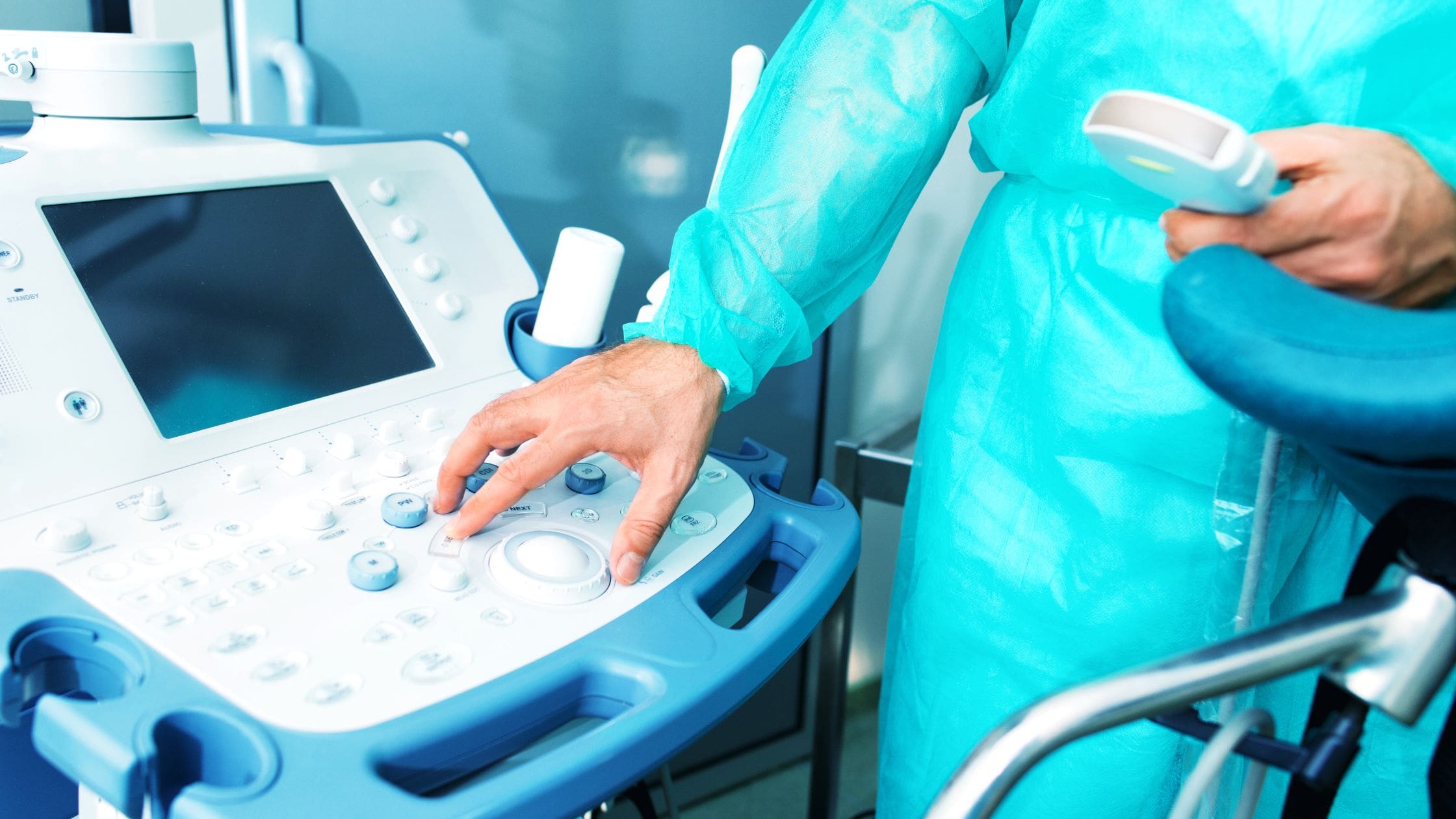
Ultrasound is safe and painless and produces images of the inside of the body using sound waves. The imaging, also called ultrasound scanning or sonography, involves the use of a transducer (probe) & specialised sound conducting gel placed directly on the skin.
High-frequency sound waves are transmitted from the probe through the gel into the body. The transducer collects the sounds that bounce back and a computer then uses those sound waves to create an image. These examinations do not use ionizing radiation (as used in x-rays), thus there is no radiation exposure to the patient. In addition, ultrasound images are captured in real-time, they can show the structure and movement of the body’s internal organs, as well as blood flowing through blood vessels.
wear loose-fitting clothing for your ultrasound exam. You may need to remove all clothing & jewelry in the area to be examined. Furthermore, you may also be asked to wear a gown during the procedure.
Preparation for the procedure will depend on the type of examination you will have. For some scans your doctor may instruct you not to eat or drink for up to 12 hours before your appointment. For others you may be asked to drink up to six glasses of water two hours prior to your exam and avoid urinating so that your bladder is full when the scan begins.
The following situations can benefit from an ultrasound examination:
Directional Doppler is used to measure velocity and direction of flow, useful in heart and blood vessel examinations.
Types of Doppler:
1: Directional: PW / CW/ AngioDoppler
2: Colour: Colour Doppler is superimposed on the 2D picture to indicate direction of flow turbulence, laminar flow , incompetence, short circuits etc.
3: Angio Doppler: Non directional, but pick up very low and slow flow and perfusion.
Doppler is essential to measure flow in the heart and to pick up valves that leak or does not open properly due to pathology. We also pick up openings in the septums of the hearts of babies or children that are born with short circuits like (ASD, VSD, open foramen ovale, PDA etc.) CW Doppler is used for higher flow velocities in one plane.
To evaluate blood vessels doppler is also essential. The important relevant vessels we examine are the following:
1: Leg veins (for deep vein thrombosis) after long flights or periods of immobility;
2: Leg arteries: when arteries narrow that supplies the muscles and feet of blood – these patients develop pain when they walk or climb stairs,with then would disappear in rest.
3: Abdominal Aorta: Some patients develop a widening or weakening of their main abdominal artery wall, called an aneurysm.
Abdominal Aorta Aneurism:
Carotid duplex studies:
Dr Grobbelaar now offers Bioidentical Hormone Replacement Therapy (BHRT). She helps manage hormonal imbalances, including menopause symptoms, fatigue and mood changes. Contact us to book a consultation.